Conditions I treat......... Limb reconstruction : Deformity correction with internal fixation

Conditions I treat.........               Limb reconstruction :       Deformity correction with internal fixation
Not all deformity correction requires an externally fixator. If an equivalent correction can be achieved with internal fixation than this is vastly preferable for the patient.

Hip reconstruction

In most cases of congenital longitudinal deficiency (leg shortening present at birth due to reduction in length and constituent elements), addressing any joint distortions merits equal consideration to correcting shortening of the limb.

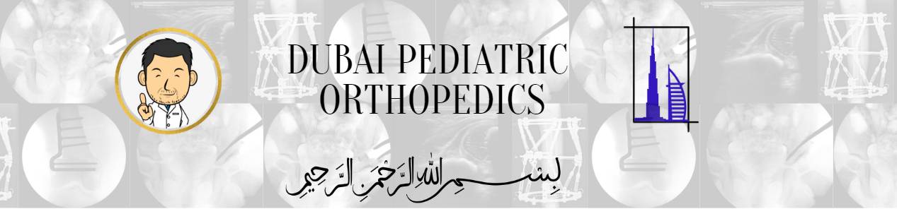
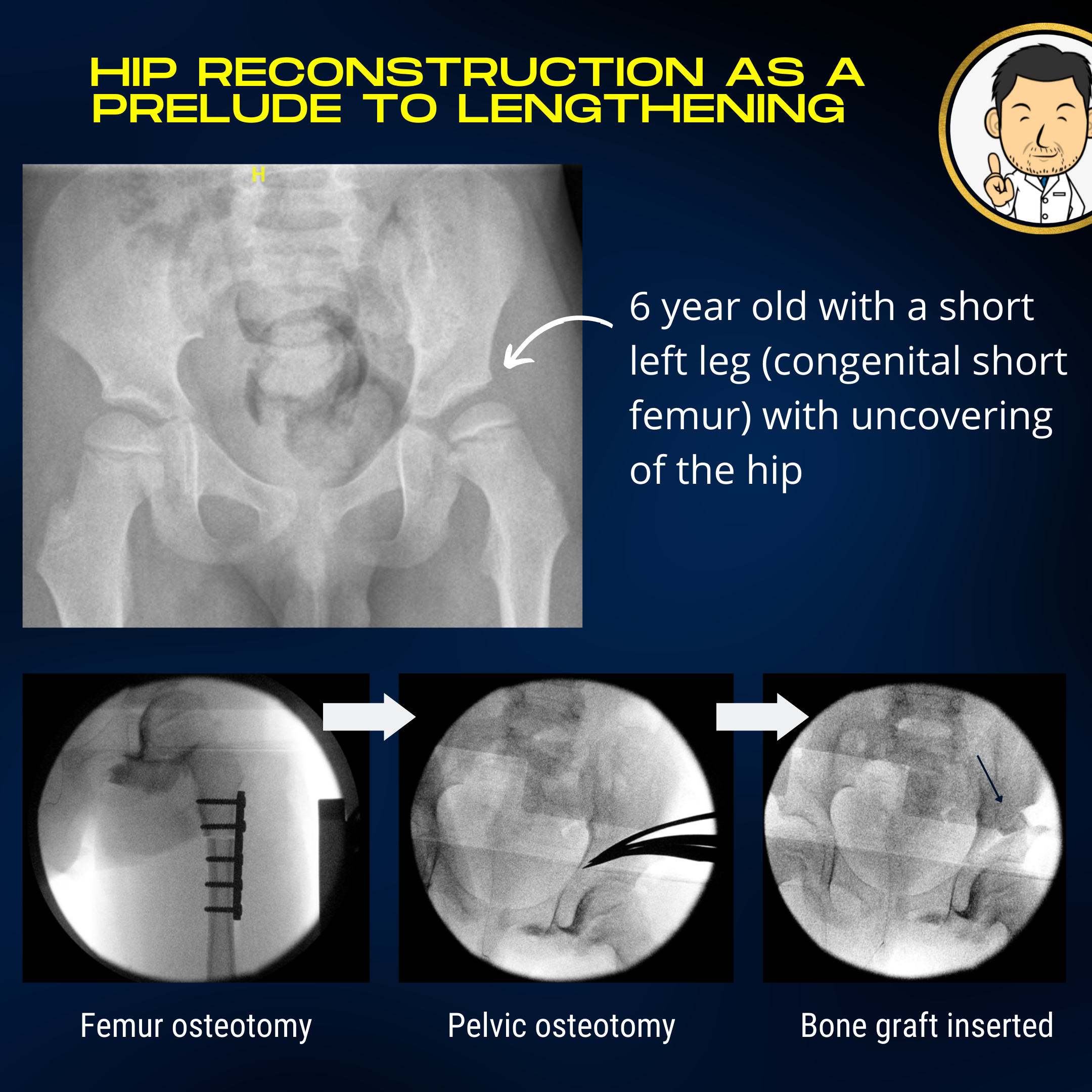
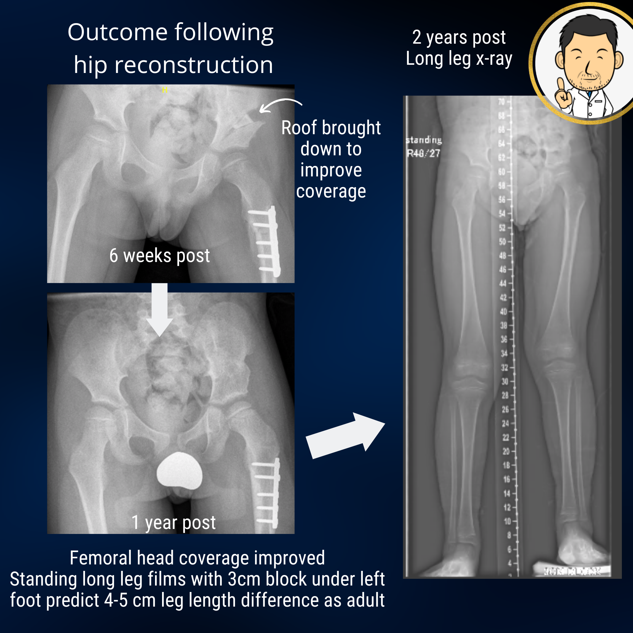
Proximal focal femoral deficiency is a congenital longitudinal deficiency predominantly affecting the femur. The mildest variant is the congenital short femur where mild femoral shortening may be accompanied by mal-alignment of the hip joint. The top end of the femur may have a "crooked hunched over" appearance and the socket may be shallow. Although, shortening in itself produces an asymmetric gait, the abnormal hip anatomy often leads to an "out-toeing" walking pattern with early fatigue due to muscle inefficiency. The socket may be shallow. If left unaddressed, this may lead to the hip dislocating if the femur is lengthened when the child is older. Correcting the alignment of the hip joint and improving socket coverage are goals that should be addressed early. The "surgical footprint" (a euphemism of the magnitude of surgery and it's impact on the patient) is much less when the hip is reconstructed early in life compared with when the child is in their teens. Early hip reconstruction improves the outcomes and risk profile of later limb lengthening surgery.

Clinical case: This 6 year old boy had a congenital short femur (VACTERL association) with uncovering of the femoral head due to a shallow socket. This can be problematic if the limb needs to be lengthened in the future as the hip could dislocate due to tension in the surrounding muscles during lengthening. Pre-emptive hip reconstruction is aimed at preparing the hip for future lengthening to minimise this risk from occurring. The rotational alignment of the femur is corrected and fixed with a plate. A bone graft taken from the femoral surgery is used to bring the socket roof down over the femoral head (black arrow - lower right image).
Clinical case cont: Outcome for the above case demonstrating improved coverage of the femoral head following hip reconstruction. Two years following surgery (right image), standing long leg films demonstrate a functional leg length difference of 3cm due to the femoral shortening. The predicted leg length difference when the child has stopped growing will be 4 to 5 cm. This child was under the 10th centile for height and mum was keen to preserve his height. Lengthening the femur can be undertaken with reduced risk of the hip dislocating due to the improved femoral head coverage.
Clinical case: Eight year old boy with right femoral shortening and coxa vara. Valgus femoral osteotomy undertaken to restore normal proximal femoral anatomy. Benefits include (1) preventing progressive deformity (further bending of neck), (2) Improve abductor lever arm (improve walking profile by mimimising "lurch" to one side), (3) improve foot progression angle (osteotomy site is rotated to bring foot in line with direction of walking) and (4) mild improvement in leg length from correction and overgrowth following osteotomy.

Knee alignment correction

In growing children, guided growth is an excellent means of tailoring growth to correct angulatory deformity around the knee. In older children, there may be insufficient remaining growth to enable such a correction. Thus, the only option is to divide and re-allign the bone to achieve the desired correction. When both angulation and length correction are required, a circular frame is often the best device. However, if the deformity is purely angular with no length considerations, correction and internal fixation with a metal plate may be the more desirable option (as stated elsewhere, "a frame is the most appriopriate option when a frame is the only option"). The clear advantage is that the fixation is all on the "inside" which is vastly less intrusive to the patient.

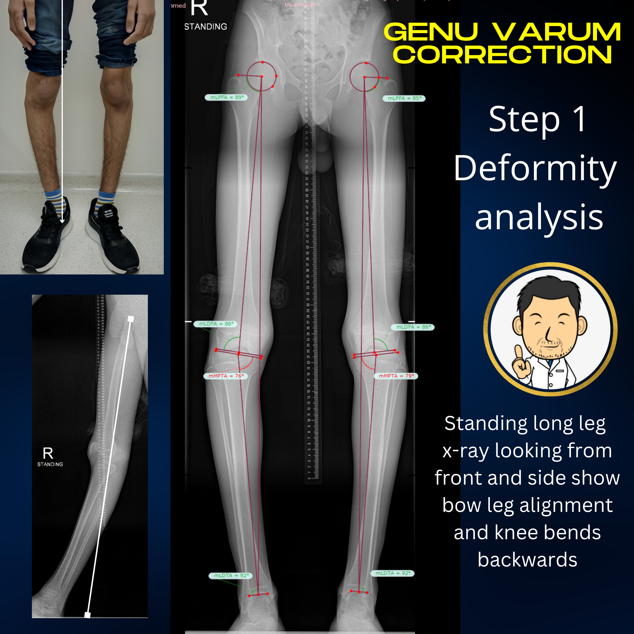
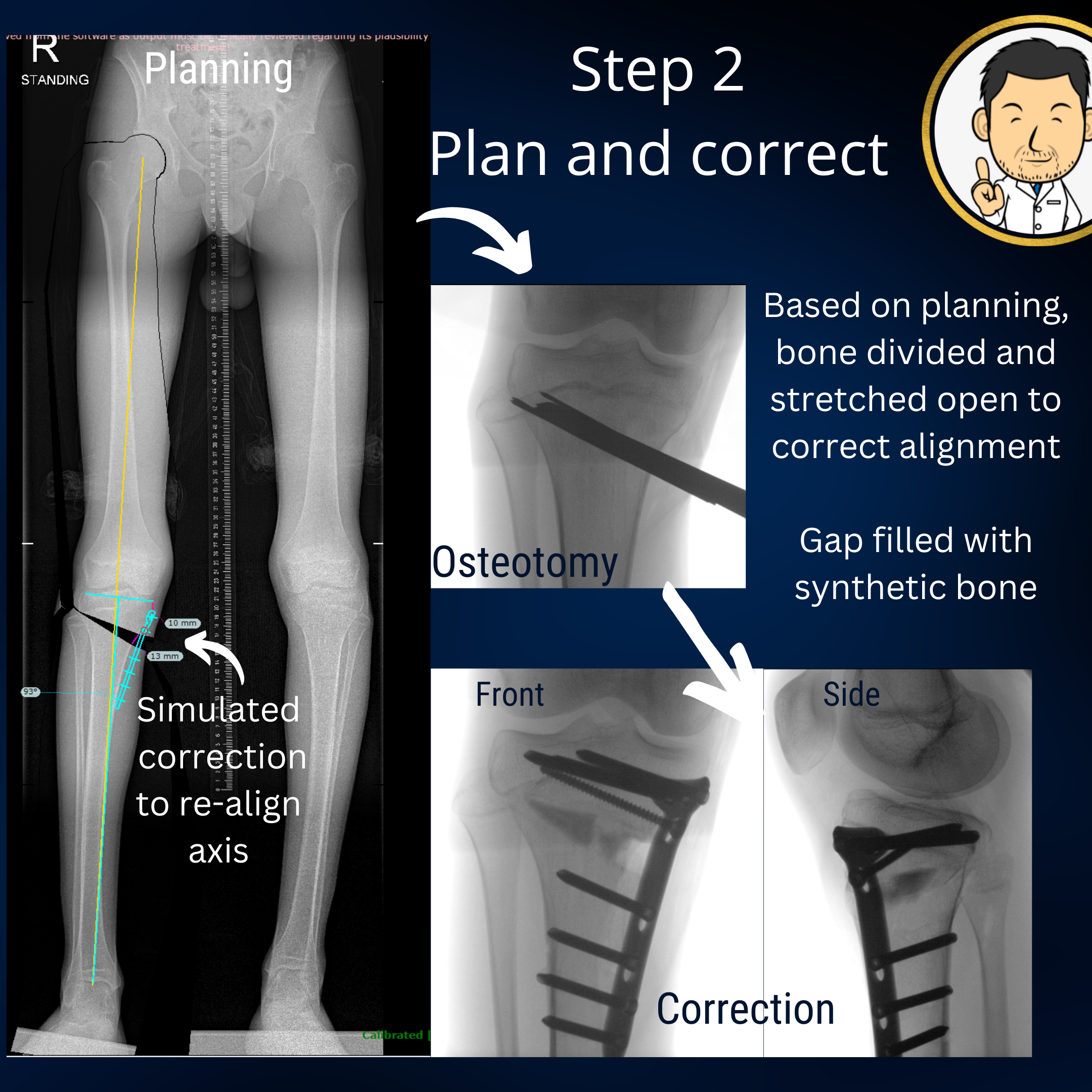
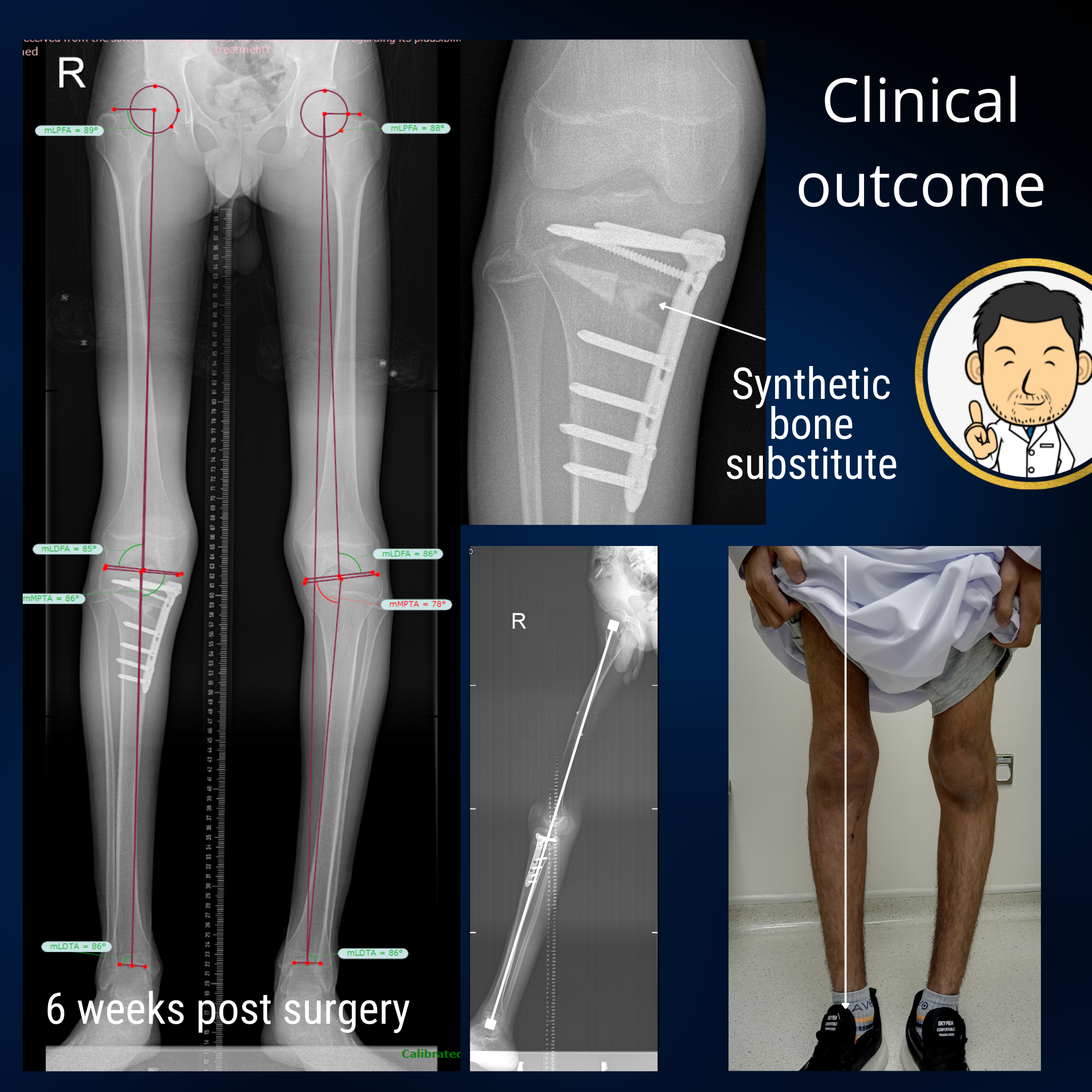
A common pattern of deformity at the knees is "bow legs" due to an oblique plane at the top of the tibia (shin bone). This is often coupled with a knee which hyper-extends (bends backwards). A carefully planned osteotomy creates an opening wedge in two planes to correct both deformities. Fixation with a plate internally is a true blessing for patients compared to treatment with an external fixation. I instruct patients to be non-weightbearing for the first 6 weeks. If all is good on the x-ray at 6 weeks, they can progress to full weight bearing thereafter. Usually I do one leg at a time so the patient can remain relatively mobile throughout treatment.

Clinical case: This 16 year old boy presented with asymmetric genu varum (bow legs). The long leg films (middle image) show theat there is mechanical axis deviation. The normal mechanical axis from the centre of the hip to the centre of the ankle passes through the centre of the knee. In this patient, the mechanical axis passes on the inside of the knee. Analysis of the joint plane obliquity suggests that the bow leg alignment is due to deformity in the top of the shin bones. The standing long leg film taken from the side (lower left image) shows that the knee bends backwards (hyper-extends) when fully straight. This is a common association - knee varus and hyper-extension.
Clinical case cont: The standing long leg x-ray is used to plan corrcetive surgery to restore the mechanical axis to the centre of the knee. The planned correction is a cut at the top of the shin bone which is wedged open and fixed with a plate. The x-rays are calibrated using the imaged ruler between the legs. This allows us to accurately measure all parameters of our planned correction i.e. where do we cut the bone (4cm from the top) and how much do we open it up by (13mm in this case). Based on the planning, the surgery is undertaken with an incomplete division of the upper end of the shin bone using osteotomes (fancy surgical chisels). The cut in the bone is wedged open to the desired 13mm. The opening is asymmetric when viewed from the side - slightly more opening at the front than at the back. This re-aligns the knee to prevent it from bending backwards when extended straight. Synthetic bone graft is wedged into the space to encourage bone growth and the correction is held with a metal plate and screws.
Clinical case cont: At 6 weeks following surgery, the patient is seen in clinic with a standing long leg x-ray. We can see that the mechanical axis from the centre of the hip to the centre of the ankle passes directly through the centre of the knee as intended. An x-ray viewed from the side (bottom middle image) demonstrates that the leg no longer bends backwards when fully straight. Clinically, the patient themself notices that their leg is now straight (bottom right image). The other leg is corrected at a subsequent operation. Correcting one leg at a time means the patient can remain relatively mobile throughout treatment.

Ankle correction

Generally the ankle is a lot more permissive for angular deformity than joints upstream. However, occasionally a situation demands that an angular correction needs to be undertaken. If there is no lengthening required, then correction with internal fixation may be feasible.

Rotational correction

The rotational alignment of the lower limbs has a broad spectrum encompassing normality. A deformity has to be well and truly two standard deviations away from normal (a statistical expression of unarguable deviation from normal) before considering surgical correction. Despite their infrequence, such cases do occur. For example, the child with external tibial torsion, out-toeing gait and marked flat feet where correction with growth is not a reasonable expectation. Or the 12 year old child with clown like appearance of moderate to severe in-toeing due to internal tibial torsion. In such cases with severe deformity and functional impairment where the capacity for natural correction has been exhausted, there is a role for surgical correction. However, to put things in context, 5 years of independent practice and thousands of patients later, I have only done this operation in a few children. Troublesome rotational deformities are more commonly encountered in children with cerebral palsy. Here the rotational deformity is but one element of a far more complex deformity pattern which needs to be corrected.